
click into different sections:
Timeline
-
2014 Discovery, Secondary structure[1]
-
2014 Crystal structure of P1-type[2]
-
2014 Crystal structure of P1-type [3]
-
2014 Crystal structure of P3-type [4]
-
2016 Application:Twister ribozymes as highly versatile expression platforms for artificial riboswitches[7]
-
2016 Active State of the Twister Ribozyme in Solution Predicted from Molecular Simulation[9]
-
2017 Chemical Mechanism[11]
-
2017 Mechanistic Debris Generated by Twister Ribozymes[12]
-
2021 The function of twister ribozyme variants in non-LTR retrotransposition[19]
Description
The twister ribozyme is a catalytic RNA structure capable of self-cleavage. The nucleolytic activity of this ribozyme has been demonstrated both in vivo and in vitro and has one of the fastest catalytic rates of naturally occurring ribozymes with similar function. The twister ribozyme is considered to be a member of the small self-cleaving ribozyme family which includes the hammerhead, hairpin, hepatitis delta virus (HDV), Varkud satellite (VS), and GlmS ribozymes.
Structure and mechanism
2D representation
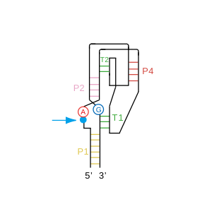
Secondary structure of the twister ribozyme. The general acid and general base are shown in red and blue respectively.
|
3D visualisation
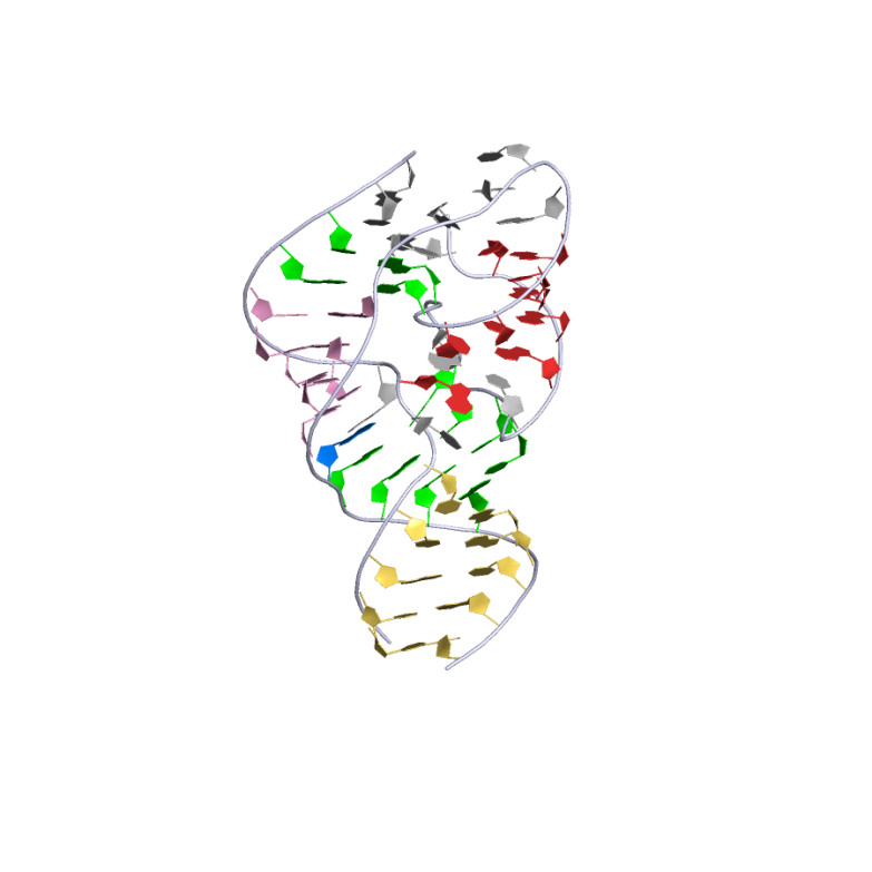
Crystal structure of the twister ribozyme. This representation was generated from PDB ID: 40JI at 2.34 Å resolution. Helices are differentiated by color. T1 and T2 are the two long-range tertiary interactions.
 |
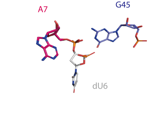
The active centre of the twister ribozyme. An O2' atom modeled onto dU6 deviates ~90° from the in-line orientation of the O2' nucleophile, P and O5' leaving group that would be optimal for activity.
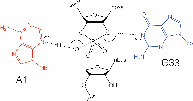
The twister ribozyme employs general acid-base catalysis, using guanine and adenine nucleobases like the hairpin and VS ribozymes, but with some important differences. The pH dependence of cleavage rate is bell shaped, corresponding to apparent pKa values of 6.9 and 9.5 [2]. Substitutions led to the assignment of the upper pKa as resulting from G33, and like hairpin and VS, G N1 is used as the general base in cleavage. An adenine is again used as general acid in the cleavage reaction, but twister differs from the other two ribozymes in two significant respects. First, it is the highly-conserved A1, immediately 3' to the cleavage site that acts as the general acid in cleavage, and an A1G mutation led to a 104-fold loss in activity. An A1 N7C atomic mutation led to a ribozyme in which the lower pKa was shifted up by 1.5 units. In its position adjacent to the scissile phosphate it is sterically impossible to employ its N1 in proton transfer. So unusually it uses the much more acidic N3 (pKa ~ 1.5) as the general acid in the cleavage reaction [6]. However, its apparent pKa is raised by formation of two hydrogen bonds from A1 N6 to negatively-charged backbone phosphate groups. The role of A1 N3 was confirmed by atomic mutation, when it was found that the activity of A1 N3C twister ribozyme was virtually undetectable while A1 N1C twister ribozyme actually became faster. The twister ribozyme has probably the best understood catalytic mechanism at the present time. The observed rate enhancement arises from four contributions [6] : References[1] A widespread self-cleaving ribozyme class is revealed by bioinformatics. [2] Crystal structure and mechanistic investigation of the twister ribozyme. [3] In-line alignment and Mg(2)(+) coordination at the cleavage site of the env22 twister ribozyme. [4] Structural basis for the fast self-cleavage reaction catalyzed by the twister ribozyme. [5] A Mini-Twister Variant and Impact of Residues/Cations on the Phosphodiester Cleavage of this Ribozyme Class. [6] The Novel Chemical Mechanism of the Twister Ribozyme. [7] Twister ribozymes as highly versatile expression platforms for artificial riboswitches. [8] High-Throughput Mutational Analysis of a Twister Ribozyme. [9] Ribozyme Catalysis with a Twist: Active State of the Twister Ribozyme in Solution Predicted from Molecular Simulation. [10] Pseudoknot Formation Seeds the Twister Ribozyme Cleavage Reaction Coordinate. [11] Unwinding the twister ribozyme: from structure to mechanism. [12] Mechanistic Debris Generated by Twister Ribozymes. [13] Metals induce transient folding and activation of the twister ribozyme. [14] Cellular Small Molecules Contribute to Twister Ribozyme Catalysis. [15] Cleaning Up Mechanistic Debris Generated by Twister Ribozymes Using Computational RNA Enzymology. [16] Classification of the nucleolytic ribozymes based upon catalytic mechanism. [17] Highly efficient expression of circular RNA aptamers in cells using autocatalytic transcripts. [18] Light-controlled twister ribozyme with single-molecule detection resolves RNA function in time and space. [19] The function of twister ribozyme variants in non-LTR retrotransposition in Schistosoma mansoni. |